G.SAI VIDYA ,IV MBBS.
We have been assigned to elog our internal assessment questions where the goal is to achieve learning driven through searchable information, particularly with regard to making original contributions and avoiding plagiarism from the searched work.
Here is my attempt of doing so.Our question paper has been prepared as to assess our ability in our clinical postings regarding " Patient clinical data analysis" to develop our competancy in comprehending clinical data including history, clinical finding, investigations and come up with a diagnosis and treatment plan.
NOTE: All the questions in the below question paper had been some of cases in our medical college in this past month.
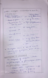
1)DEFINE BONE DENSITY, HOW IS IT MEASURED? WHAT ARE THE CAUSES, CLINICAL FEATURES, DIAGNOSIS AND MANAGEMENT OF OSTEOPOROSIS ?
Bone density:
Also called BMD, bone mass, and bone mineral density.
A measure of the amount of minerals (mostly calcium and phosphorous) contained in a certain volume of bone. Bone density measurements are used to diagnose osteoporosis (a condition marked by decreased bone mass), to see how well osteoporosis treatments are working.
A bone density test is used to measure bone mineral content and density. It may be done using X-rays, dual-energy X-ray absorptiometry (DEXA or DXA)
MEASURING BMD:
• Z score: the number of standard deviations above or below the mean for the patient's age, sex and ethnicity.
• T score: the number of standard deviations above or below the mean for a healthy 30-year-old adult of the same sex and ethnicity as the patient.
2)DEFINE MYXOEDEMA COMA? DESCRIBE ITS CLINICAL FEATURES, DIAGNOSIS AND TREATMENT.
3)WHAT IS THE DIAGNOSTIC APPROACH OF YOUNG ONSET HYPERTENSION AND ITS TREATMENT?
Young-onset hypertension is a common condition that increases all-cause mortality and results in subclinical organ damage early in its natural history. Epidemiological studies suggest that early-life factors are important and should be addressed by public health policies to reduce cardiovascular disease later in life.
Referral of selected patients to secondary care (is suggested as it permits more detailed assessment, evaluation of subclinical organ damage, and investigation of secondary causes of hypertension. This can allow more personalized management given the limitations of current risk-based approaches in this population. Early intervention with medication in young, mild hypertensives (blood pressure, >140/90 mm Hg) free of cardiovascular disease could be considered following a 1-year trial of lifestyle modification.
4)HOW DO YOU CLINICALLY LOCALISE THE ANATOMICAL LEVEL OF LESION IN SPINAL CORD DISEASES


In cases of a lower motor neuron type weakness, there is early muscle wasting, fasciculations, hypotonia, hyporeflexia, and a normal plantar response. On the other hand, the upper motor neuron type of weakness is characterized by normal muscle bulk, hypertonia, hyperreflexia, clonus, and an extensor plantar response (positive Babinski’s sign). Furthermore, the preservation of deep tendon reflexes distinguishes myopathy from neuropathy.
Lower motor neuron type weakness can result from any pathology of anterior horn cells, spinal nerve roots, plexus, or peripheral nerves.
An upper motor neuron paraplegia can result from myelopathy involving the thoracic spinal cord, while cervical myelopathy would result in quadriplegia. The uppermost dermatomal level of accompanying sensory loss can further localize the lesion to the corresponding spinal segment.
Myelopathy is further classified into compressive and non-compressive myelopathy. An example of non-compressive myelopathy is transverse myelitis that can be partial or complete. Compressive myelopathy can result from compression from the outside like a vertebral fracture, vertebral metastasis or subdural abscess or hematoma, or from the inside with conditions such as syringomyelia
5) CAUSES, DIAGNOSIS AND TREATMENT OF ATRIAL FIBRILLATION?
6)DESCRIBE ABOUT MEGALOBLASTIC ANEMIA
7)WHAT ARE THE CAUSES PATHOGENESIS AND DDs OF ASCITES?
DDs for ascites:
• Acute Liver Failure
• Biliary Disease
• Budd-Chiari Syndrome
• Cirrhosis
• Dilated Cardiomyopathy
• Familial Mediterranean Fever
• Hepatocellular Adenoma (Hepatic Adenoma)
• Hepatorenal Syndrome
• Nephrotic Syndrome
• Portal Hypertension
• Primary Biliary Cholangitis (Primary Biliary Cirrhosis)
• Protein-Losing Enteropathy
• Restrictive Cardiomyopathy
• Viral Hepatitis
8)APPROACH TO ACUTE PANCREATITIS
9)MENTION THE DIFFERENCE BETWEEN UMN AND LMN LESION?
10)INDICATIONS OF HEMODIALYSIS
Indications of hemodialysis:
-Refractory fluid overload
-hyperkalemia(plasma potassium concentration >6.5 meq/L)
-metabolic acidosis(pH less than 7.1)
-azotemia(BUN greater than 80 to 100 mg/dL)
-signs of uremia ,such as pericarditis ,neuropathy etc
11) WHAT IS THE ROLE OF SUCRALFATE IN EROSIVE GASTRITIS
12)RENAL MANIFESTATIONS OF SNAKE BITE
clinical renal manifestations vary from mild proteinuria, haematuria, pigmenturia to acute renal failure. Bites by haemotoxic snakes and myotoxic snakes are the common causes of renal involvement especially acute renal failure.
Mesangiolysis, glomerulonephritis, vasculitis, tubular necrosis, interstitial nephritis and cortical necrosis. Tubular necrosis is an important pathological counterpart of acute renal failure.
13)CAUSES OF PORTAL HYPERTENSION
Prehepatic:
-Congenital stenosis of portal vein
-External compression of portal vein
-Arterio portal fistula
Hepatic:
-Cirrhosis
-Congenital hepatic fibrosis
-Hepatic portal sclerosis
-Nodular regenerative hyperplasia
-Venoocclusive disease
-Granulomatous disease
Post hepatic:
-Hepatic vein thrombosis
-Congenital malformations and thrombosis of IVC
-Constrictive pericarditis
14)DOWN’S SYNDROME
15)Renal manifestations of Post streptococcal Glomerulonephritis
16)CAUSES OF CERVICAL MYELOPATHY


Comments
Post a Comment